

Blog after blog, we discuss the pelvic floor and the musculoskeletal system contained within this region of the body. In case any of you have wondered, “Hmm…what exactly does she mean when she talks about the pelvic floor?…” today is your lucky day! Because today, dear reader, we will discuss some of basic anatomy and physiology of the area we refer to as the pelvic floor.
The pelvic floor is a truly incredible system. Several noteworthy anatomical landmarks include:
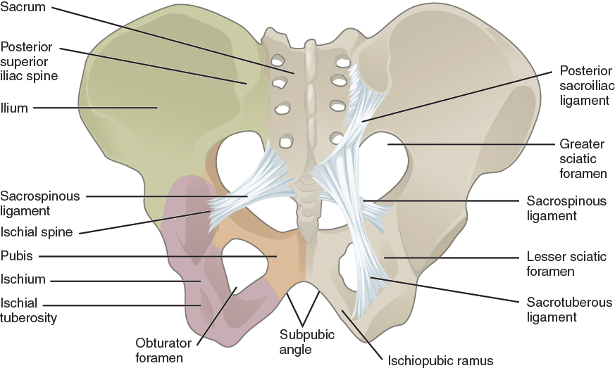
- Hip bone- comprised of a left and right ilium, ischium, and pubic bone
- Pubic symphysis- this cartilaginous joint is located at the junction of the left and right superior rami of the pubic bones. The bladder is located behind it and the clitoris and external genitalia are located below it. This joint acts like a cushion and serves as a shock absorber during walking. It also allows for pelvic expansion during labor and delivery. The pubic symphysis has a small degree of superior/inferior mobility (movement up or down) as well as the ability to separate slightly, as required during pregnancy to accommodate for the growing fetus. The space in between the two pubic bones is usually 4-5 mm, however this can increase 2-3 mm during pregnancy. Diastasis of the symphysis pubis, also known as pubic symphysis separation, is a dysfunctional excessive widening of the pubic symphysis joint during pregnancy or after delivery.
- Sacrum- This bone looks like an upside down triangle and it rests at the base of the spine. It consists of five smaller sacral bones (or vertebrae) which fuse between the ages of 18 and 30. It connects to the fifth lumbar vertebrae which sits above it, the coccyx which sits below it, and the left and right ilium bones of the hip. Several important muscles which allow for movement of the legs, including the gluteus maximus, iliacus, and piriformis, originate from the sacrum and are attached to this bone.
- Coccyx- This bone, colloquially referred to as the tailbone, is a small, often overlooked anatomical landmark that sits below the lumbar spine and sacrum. Despite its tiny size, it is extremely clinically significant. Think of it as the Grand Central Station of your pelvic floor, so to speak. The coccyx serves as the attachment site of the gluteus maximus and levator ani muscles (which include the coccygeus, iliococcygeus, and pubococcygeus muscles) and ligaments (including the anterior, posterior, and lateral sacrococcygeal, sacrotuberous, and sacrospinous ligaments). Injury to the coccyx can affect the aforementioned muscles and/or ligaments. Conversely, injury to the muscles and/or ligaments can affect coccyx alignment. Symptoms of coccyx dysfunction include coccyx pain (referred to as coccydynia), pain with defecation, pain with intercourse, pain with prolonged sitting, pain with transitional movements (such as sit to stand), coccyx pain, low back pain, and even neck pain
- Ischial tuberosities- these prominent bumps (or protuberances) are located on the left and right ischium bones, and they are commonly referred to as the sitz bones. Most people bear weight onto the ischial tuberosities while sitting. The hamstrings muscles and sacrotuberous ligaments attach to the ischial tuberosities.
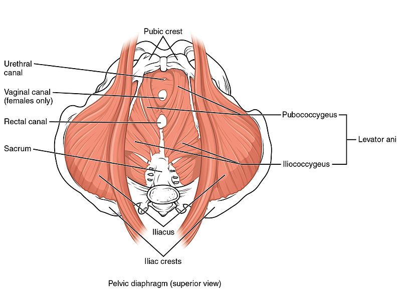
Four layers of muscular tissue (aka diaphragms) exist within the pelvic floor:
- Superficial perineal space, the area overlying the pelvic outlet, which includes the genitalia, urogenital triangle, anal triangle, perineal body
- Urogenital diaphragm- contains deep transverse perineal muscle and sphincter urethrae (contributes to voluntary control of voiding)
- Pelvic diaphragm (aka levator ani muscles)- this is the deepest layer of striated muscle. The two primary functions of this layer of muscles is to elevate the pelvic floor and to resist intra-abdominal pressure. It consists of four muscles (one on each side), the pubococcygeus, the puborectalis (controls descent of feces, creates anorectal junction/angulation), the iliococcygeus, and the coccygeus.
- Smooth muscle diaphragm- this contains the internal urethral sphincter and the external urethral sphincter
In addition, there are three openings within the pelvic floor. From front to back, the three openings are:
- The urethra, which connects to the bladder and is involved with elimination of urine.
- The vagina, which connects to the uterus and is the opening through which the fetus is delivered at the end of pregnancy.
- The anus, which connects to the rectum and is involved with elimination of stool.
The area located between the vagina and the anus is referred to the perineal body.
Hopefully, this little anatomy lesson has given you a better frame of reference for any discussion about women’s health. Understanding one’s body and how it functions will hopefully lead to a greater appreciation of the daily miracles that occur as well as lead to quicker diagnosis and treatment of dysfunction.